New Ways to Identify Chronic Pain
This project seeks to develop and validate a quantitative biomarker that enables the differentiation between normal physiological processes in soft tissues such as muscle, connective tissue, nerve, blood vessels and extracellular fluid, and pathological processes in these tissues that may be associated with myofascial pain syndrome.
This project aims to create and test a reliable biomarker to measure changes in soft tissues, such as muscles, nerves, connective tissue, blood vessels, and fluid. The goal is to distinguish between normal tissue behavior and changes linked to myofascial pain syndrome.
Fluorescent Nanoparticles for Deep Tissue Imaging
This project bridges the fields of medical imaging and synthetic biochemistry to create nanoscale sensors that measure and analyze tissue structure and function. Our team specializes in developing advanced fluorescent nanoparticles designed to detect and quantify nervous tissue activity and assess bioretention in cancerous tissues. Additionally, we have pioneered a versatile nanoparticle platform that enables the delivery of vaccines, contrast agents, and other therapeutic compounds, advancing both diagnostic and therapeutic capabilities in biomedical research.

Confocal fluorescence microscopy image of a neuron in the mouse hippocampus
Nanomaterials for Therapeutic and Diagnostic Applications
In collaboration with Dr. Remi Veneziano‘s lab, this project focuses on the design and development of advanced nanomaterials, including DNA scaffolds, dye aggregates, lipid nanoparticles, and liposomes for dual therapeutic and diagnostic applications. Central to these platforms is the incorporation of organic dyes, which enables functionalities such as near-infrared II (NIR-II) fluorescence and photoacoustic imaging. Among these, DNA scaffolds are particularly versatile, allowing precise control over size, shape, and surface functionalization to template dyes and targeting moieties for enhanced specificity and sensitivity. They are being developed for applications such as tumor imaging, neurosensing, deep tissue imaging, image-guided surgery, and photothermal therapy. All probes are characterized and evaluated through in vitro, ex vivo, and in vivo studies to assess their performance.
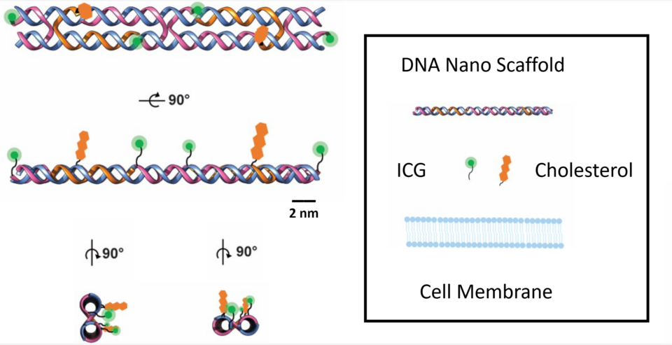
Novel DIVIN Nano-sensor: Schematic of DIVIN made in Chimera shown bound to ICG and cholesterol. The structure has been designed to have the different moieties facing the same direction
DNA-Based Near-Infrared Voltage Sensors, ACS Sensors

(Left) Illustration of fluorophore photoacoustic and fluorescence emission following light excitation; (Right) Jablonski diagram describing the energy acquisition and release pathways of fluorescence and photoacoustic imaging
NIR-II Nanoprobes: A Review of Components-Based Approaches to Next-Generation Bioimaging Probes, MDPI Bioengineering

Fluorescence Intensity Changes of A Target Hela Cell Stained with Voltage Sensing Fluovolt Dye in Response to Induced Voltage
DNA-Based Near-Infrared Voltage Sensors, ACS Sensors
Flexible and Wearable Nanocomposite-based Ultrasound Transducers
Ultrasound technology plays an essential role in diagnostic imaging and is widely used in medical applications. However, conventional ultrasound transducers face challenges related to flexibility and fabrication complexity, driving the development of innovative thin-film materials and advanced manufacturing techniques to enhance transducer performance. The goal of this project is to find novel methods of fabricating inexpensive, flexible transducer patches using laser induced graphene and flexible piezomaterial.